THE DETAIL TO KEEP
The ulcer paper appeared in 1994; its later PubMed indexing does not make it a 2006 trial.
Sections in this fieldnote
Copper-peptide research spans very different experiments: cells making connective-tissue proteins, a gel tested in diabetic ulcers, and skincare regimens used after laser resurfacing. These studies do not form a single interchangeable proof of skin rejuvenation. Each asks a different question about a particular biological setting, intervention, and measurement. The useful connection is a research pathway—from plausible cellular activity to a clinical test—not a shortcut from laboratory activity to a retail product claim.
The ulcer study offers an especially instructive example because its striking result concerns the percentage of wound area closed, not the percentage of participants healed. This analysis uses the indexed primary abstracts and an author-posted full-text transcription of the ulcer report. The latter adds important design details, although its imperfect optical character recognition leaves some table entries unsuitable for publication. The laser and cell studies remain abstract-only evidence here.
Study facts at a glance
Historical publication: The ulcer paper appeared in 1994; its later PubMed indexing does not make it a 2006 trial. Source 5 Source 2
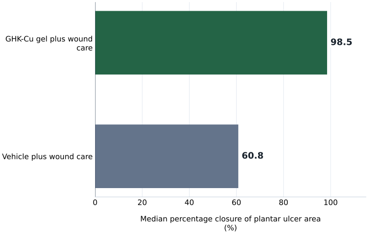
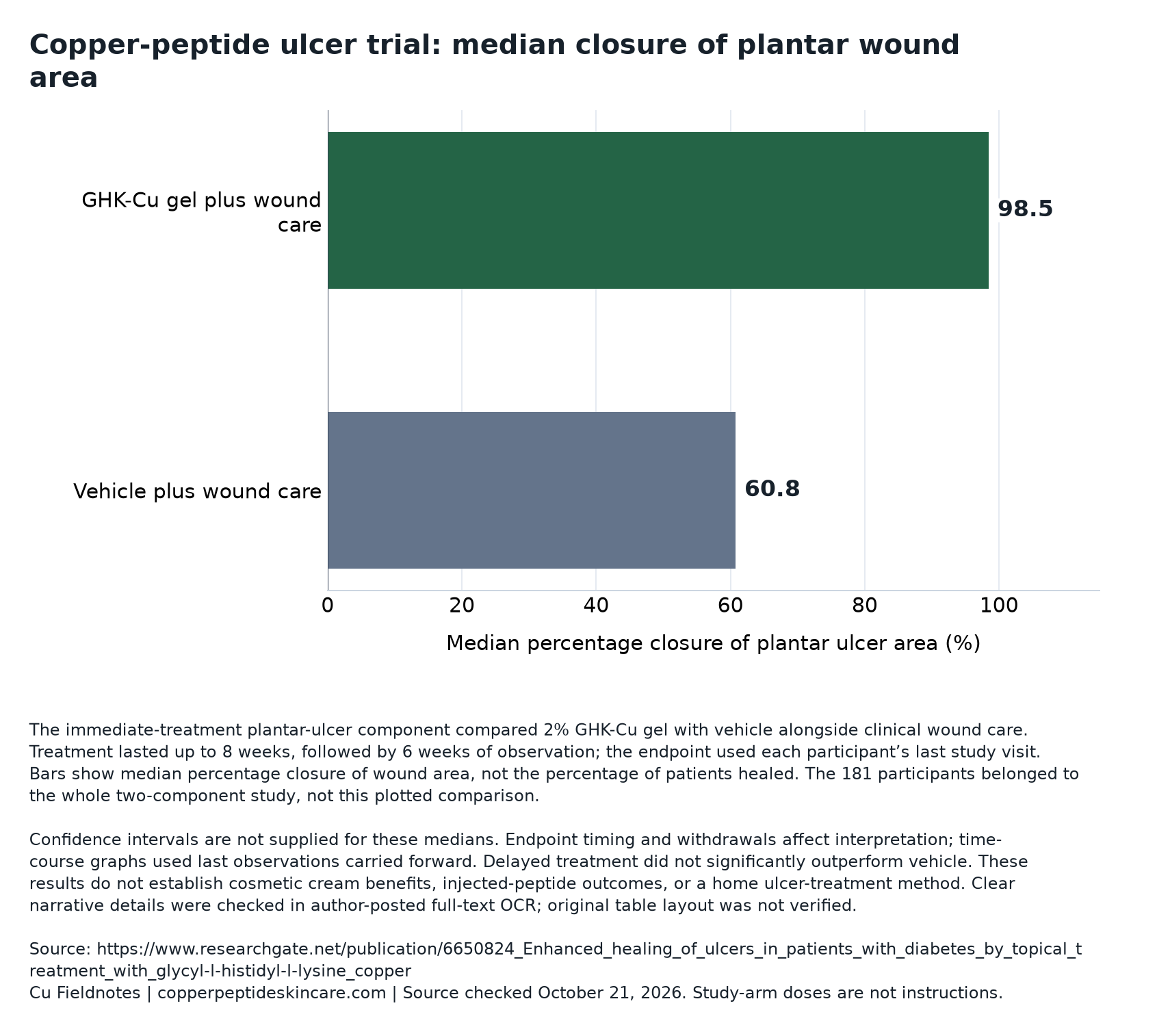
Main numerical finding: Median plantar-ulcer area closure was 98.5% with gel versus 60.8% with vehicle in the immediate-treatment component. Source 2 Source 5
Whole-study enrollment: The report enrolled 181 people; that total is not the denominator for the immediate-treatment plantar comparison. Source 5
Laboratory context: Fibroblast experiments examined collagen production and matrix-remodeling signals, not cosmetic outcomes in people. Source 3 Source 4
Different human question: The laser study separated objective skin assessments from patient-reported improvement. Source 1
What the ulcer comparison actually tested
The multicenter trial was randomized, evaluator-blinded, and vehicle-controlled. Participants had diabetic neuropathic ulcers; all groups received standardized debridement, pressure-relieving footwear, and education. Immediate treatment compared topical 2% GHK-Cu gel with vehicle for up to eight weeks, followed by six weeks of observation. A delayed component used four weeks of vehicle before eight additional weeks of vehicle, 2%, or 4% gel. These concentrations identify historical study arms, not treatment instructions.
The comparator matters as much as the active ingredient. A vehicle is the formulation without the tested active component. Comparing active gel with its vehicle, while providing clinical care to both groups, asks whether adding that formulation improves outcomes beyond the shared program. It does not compare copper peptide with doing nothing, and it does not isolate the value of every element of wound care.
Interpretation: the most defensible claim has several qualifiers attached—this topical formulation, in this clinical population, alongside this care program. Removing those qualifiers makes the claim broader than the experiment. The distinction also explains why a wound-gel study belongs beside, rather than inside, a general assessment of copper peptides and human skin evidence.
Read the underlying chart data: download CSV or download the source-labeled chart.
Area reduction is not a patient healing rate
The abstract reports median area closure of 98.5% versus 60.8%, with P < .05. Subtracting these summaries gives a 37.7-percentage-point difference in median area closure. That is not a 37.7-point increase in the probability of complete healing. A median describes the middle of an ordered set of measurements; it is not a count of people crossing a clinical finish line.
Illustrative reasoning: imagine one ulcer initially covering 200 square millimeters. A 60% reduction would leave 80 square millimeters open; a 98% reduction would leave four. Both represent substantial shrinkage, but neither calculation establishes complete closure. These hypothetical dimensions explain the endpoint only; they are not reconstructed participant data.
A separate categorical endpoint asks how many wounds cross a specified threshold. That question needs a numerator and denominator, not just a median. The report also examined near-complete closure, but the transcription's categorical table is not sufficiently secure to publish its proportions. Keeping the two endpoints separate prevents the headline number from silently becoming a success rate.
Three wound measurements answer different questions
| Measurement | What it describes | What it does not establish |
|---|---|---|
| Median percentage area closure | The middle percentage reduction among measured ulcers | The percentage of participants completely healed |
| Categorical closure threshold | The fraction crossing a defined area-reduction boundary | Durable healing unless durability is separately assessed |
| Normalized closure rate | Movement of the wound edge over time, adjusted for geometry | A multiplier for the number of people healed |
Why an enlarging wound produces a negative number
For the larger-ulcer subgroup, the abstract reports median closure of 89.2% with gel versus −10.3% with vehicle, with P < .01. Its size definition uses greater than 100 square millimeters; the body transcription uses a boundary including 100. The transcription also describes 10.4% expansion rather than 10.3%. Those small discrepancies remain unresolved, so the abstract's figures should retain that attribution.
The negative sign has a straightforward meaning. Percentage closure can be written as starting area minus later area, divided by starting area, multiplied by 100. In an illustrative ulcer growing from 100 to 110 square millimeters, this produces −10%. There is no such thing as a negative percentage of patients healing; the number describes expansion of an individual wound's area.
This scale is asymmetric. Closure stops at 100%, but enlargement has no corresponding lower boundary: doubling in area gives −100%, and tripling gives −200%. Consequently, a few greatly enlarged wounds can pull an arithmetic mean downward much more than nearly closed wounds can pull it upward. A median reduces that leverage, although it also hides how widely individual experiences differ.
The endpoint clock was not identical for everyone
Weekly tracings supplied area measurements. The end-of-study endpoint used each participant's last visit, whether departure followed closure, an adverse event, or another reason; longitudinal graphs carried the last value forward. All enrolled participants were included in analyses, including four with only a prestudy evaluation. Thus, the endpoint should not be described as a uniform 14-week measurement.
Why does that matter? Consider two hypothetical participants: one leaves early after closure, while another leaves early with an unresolved ulcer. Their last measurements can both enter an endpoint analysis, but they represent different amounts of observed time. Including everyone preserves more of the original comparison; it does not magically supply the later outcomes that were never measured.
Carrying a value forward is an assumption about missing observations, not a new observation. It holds the recorded state constant after departure. A modern sensitivity analysis could ask whether conclusions change under plausible improvement or deterioration after missing visits. Without such checks, readers should distinguish the measured last-visit result from claims about sustained closure or recurrence.
Timing findings do not establish a mechanism
The delayed gel comparisons did not show statistically significant improvement over vehicle. This makes timing a substantive part of the clinical result rather than an incidental scheduling detail. It also prevents the immediate-treatment finding from being generalized across every active arm.
A timing difference can generate hypotheses without deciding among them. Treatment access to tissue, the changing wound environment, and the trajectory following a procedure could all be candidates for investigation. Demonstrating one would require measurements specifically designed to distinguish it from alternatives. Merely observing different outcomes after different schedules cannot establish which biological step caused the difference.
There is a second analytical issue: before-and-after comparisons can confuse treatment effects with a naturally changing recovery curve. If a wound's early trajectory differs from its later trajectory, using its early period as a baseline does not automatically provide a valid untreated counterfactual. The methodological lesson is to preserve the distinction between a concurrent comparator and a participant's own earlier course.
A faster edge is not three times as many healed wounds
The ulcer abstract describes a threefold faster closure rate. The full report identifies a normalized linear-edge measure in millimeters per day. This is a geometric rate, not a risk ratio for healing. It should not be translated into three times as many people healed or a promise that treatment takes one-third as long.
Illustrative reasoning: area and edge movement are related but not interchangeable. For a perfectly circular wound, area equals π multiplied by radius squared. Moving the boundary inward by one millimeter removes a different amount of area from a large circle than from a small one. A normalized edge measure tries to make comparisons less dependent on starting size and shape.
Even then, a summarized rate does not imply constant movement. Real trajectories may accelerate, stall, or reverse. A better future report would present both rate and clinically meaningful completion outcomes, with uncertainty intervals and clearly specified follow-up. Those measures complement one another: geometry describes progress, while completion and durability describe whether an important clinical state was reached and maintained.
Infection findings need their own denominator
Reported infections were 7% with gel versus 34% with vehicle in immediately treated plantar ulcers, with P < .05. Overall adverse-event rates did not differ significantly. Infection unresponsive after a week of therapy triggered withdrawal. These findings are not equivalent to a blanket declaration of safety.
The infection comparison is an absolute difference of 27 percentage points in that specified subset. It is not a whole-trial infection estimate, and it does not demonstrate infection prevention in other wounds. Without verified subgroup counts and confidence intervals, a reader cannot adequately reconstruct precision or judge how sensitive the estimate is to a small number of events.
A nonsignificant overall comparison also leaves several possibilities open, including similar event rates or insufficient precision to distinguish them. Withdrawal rules connect safety with outcome interpretation because subsequent wound status may become unobserved. The clinical setting should remain explicit: historical supervised research is not a basis for self-treating diabetic ulcers with cosmetics. The open-wound evidence boundary addresses that distinction.
Cell experiments suggest remodeling, not simply more collagen
In fibroblast cultures, GHK-Cu stimulated collagen synthesis independently of cell-number changes. Reported activity began between 10⁻¹² and 10⁻¹¹ molar and peaked at 10⁻⁹ molar. A separate culture study found increased MMP-2 protein and messenger RNA, alongside increased secretion of TIMP-1 and TIMP-2. Copper ions reproduced the MMP-2 response; GHK alone did not.
Fibroblasts are connective-tissue cells. MMPs are enzymes involved in matrix breakdown, while TIMPs inhibit metalloproteinases. Seeing both production and regulatory signals makes “remodeling” a more useful interpretation than a simple accumulation story. However, measurements in culture do not reveal the net balance of tissue building and breakdown in a person, much less whether that balance produces a visible cosmetic benefit.
An experimental concentration is the amount presented under laboratory conditions, not a validated concentration for a bottle. Translation requires information about chemical form, availability, exposure, and tissue access. Matching a label number to a culture concentration skips those steps. The guide to copper-peptide percentages and labels explains why a percentage alone cannot supply that bridge.
The research settings test different links in the evidence chain
| Setting | Measured question | Remaining translation question |
|---|---|---|
| Collagen fibroblast culture | Does collagen synthesis change under controlled exposure? | Does a specific human formulation produce a meaningful tissue outcome? |
| Matrix-remodeling fibroblast culture | Do enzyme and inhibitor signals change? | What is the net effect in living tissue? |
| Diabetic-ulcer trial | Does adding a topical gel improve specified wound outcomes? | How reproducible and durable is the clinical effect? |
| Post-laser skincare trial | Do recovery assessments or patient reports differ between regimens? | Would findings apply to routine use on intact skin? |
Source 3 Source 4 Source 2 Source 5 Source 1
The laser study supplies a different clinical test
In the randomized post-laser study, 13 patients completed treatment; enrollment, arm sizes, and attrition details were unreported in the abstract. Objective assessments found no significant between-group advantage for redness resolution, wrinkles, or overall skin quality. Both groups improved after resurfacing. A patient questionnaire favored the GHK-Cu regimen for perceived overall skin quality, with P = .04.
These observations distinguish change over time from the added effect of a regimen. Improvement in both groups does not show that both skincare regimens caused the improvement; both groups also underwent laser treatment. Conversely, a nonsignificant between-group result does not mean that values stayed unchanged, nor does a small study establish equivalence.
Subjective and objective outcomes can legitimately answer different questions. Satisfaction may matter to participants without proving a measurable structural benefit. The abstract supplies no effect-size confidence intervals or adverse-event counts. The companion analysis of the laser-resurfacing trial explores those endpoint distinctions. Neither clinical study directly answers whether a routine facial cream improves intact skin.
What would strengthen the clinical case
The ulcer report shows ProCyte involvement through corporate affiliations, the identified study product, and acknowledgment of its development team; that is not a verified formal funding statement. Commercial involvement is relevant context, not a numerical correction to the findings. The stronger response is to seek transparent methods, independently reproducible results, and access to interpretable outcome tables.
Important next questions include a prespecified main endpoint, confidence intervals, dependable subgroup denominators, durable closure, and missing-data sensitivity analyses. Multiple endpoints and subsets also raise the question of which comparisons were planned and how multiplicity was handled. Statistical significance alone cannot establish the size, reliability, or practical importance of an effect.
Editorial interpretation: purchasing and service-contract questions belong on a separate track from clinical efficacy. A cream, serum, compounded preparation, oral product, or systemic injection does not inherit the gel's evidence. The cream-versus-serum formulation guide helps frame that distinction. For documentary terms rather than inferred outcomes, readers can consult the AgelessRx review or Defy Medical review; neither link establishes access to the studied treatment.
Questions about the evidence
Does nearly complete area closure mean the wound was healed?
Not necessarily. Area reduction, complete closure, and durable closure are distinct outcomes. A small remaining opening still differs from no opening, and an initially closed wound may require later observation to establish durability. A median area result cannot answer all three questions.
Why not calculate a number needed to treat from these findings?
That calculation requires a reliable absolute difference in a clearly defined patient-level event over a specified period. Median area closure is not such an event. The available transcription also does not securely establish the categorical table values needed for a defensible calculation.
Do the cell findings explain the ulcer benefit?
They provide biological hypotheses, not a demonstrated causal pathway in trial participants. Establishing mediation would require evidence that treatment changed the proposed biological process in people and that this change accounted for the clinical outcome. Parallel laboratory and clinical findings do not establish that connection by themselves.
Is the lack of a significant delayed-treatment benefit proof that delayed treatment has no effect?
No. A nonsignificant comparison does not prove a zero effect. It does limit affirmative claims: the immediate-treatment finding cannot simply be extended to the delayed schedules. Confidence intervals and a suitably designed replication would help distinguish an absent effect from an imprecisely estimated one.
Does a copper-peptide ingredient name make two products clinically equivalent?
No. Shared ingredient terminology does not establish matching formulation, exposure, tissue access, route, or intended use. Equivalence is a question requiring evidence about the actual products. Neither cellular activity nor success in a wound-care setting automatically validates a cosmetic or systemic preparation.
What this evidence supports
The ulcer trial supports a specific clinical signal for an immediately applied topical gel within standardized care, not a general copper-peptide healing or anti-aging claim. Its most useful lesson is methodological: identify the population, comparator, timing, endpoint, and denominator before interpreting a dramatic percentage. Cell-culture findings make further investigation reasonable, but they cannot fill gaps in clinical precision, durability, formulation equivalence, or cosmetic effectiveness.
Research access and limitations
The ulcer analysis uses an author-posted primary full-text OCR with unverified table and figure layout, alongside its indexed abstract. Ambiguous arm counts and categorical proportions are omitted. Confidence intervals are unavailable in the accessible material. The laser and laboratory analyses use indexed primary abstracts, not reviewed full protocols or supplements. These historical reports are not current prescribing information.
SOURCE NOTES
What this record draws on
- Topical copper tripeptide after CO₂ laser resurfacing (2006) ↗
Abstract reviewed: 13 completers in a post-laser setting. Objective between-group differences were not significant; patient-reported satisfaction favored GHK-Cu. Not a trial of the current reviewed products. Accessed 2026-10-21.
- Enhanced healing of ulcers in patients with diabetes by topical treatment with glycyl-l-histidyl-l-lysine copper. ↗
Indexed primary study abstract Accessed 2026-10-21.
- Stimulation of collagen synthesis in fibroblast cultures by the tripeptide-copper complex glycyl-L-histidyl-L-lysine-Cu2+. ↗
Indexed primary laboratory-study abstract Accessed 2026-10-21.
- The tripeptide-copper complex glycyl-L-histidyl-L-lysine-Cu2+ stimulates matrix metalloproteinase-2 expression by fibroblast cultures. ↗
Indexed primary laboratory-study abstract Accessed 2026-10-21.
- Mulder et al., 1994: GHK-Cu in diabetic neuropathic ulcers (author-posted full text) ↗
Author-posted primary full-text OCR; original table and figure layout not verified Accessed 2026-10-21.
Individual source access dates are recorded above. Prices, stock, labels, and directions can change. How we review.